Internal Jugular Vein Course Tributaries Relations AnatomyQA
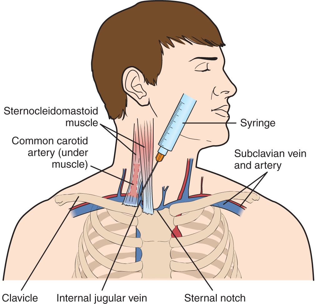
Ultrasound-guided cannulation of the internal jugular vein uses real-time (dynamic) ultrasound to guide venipuncture and a guidewire (Seldinger technique) to thread a central venous catheter through the internal jugular vein and into the superior vena cava.
/GettyImages-530309436-cf8e158016cf4dc0a81e12ecb221d1ee.jpg)
Internal Jugular Vein Anatomy, Function, and Significance
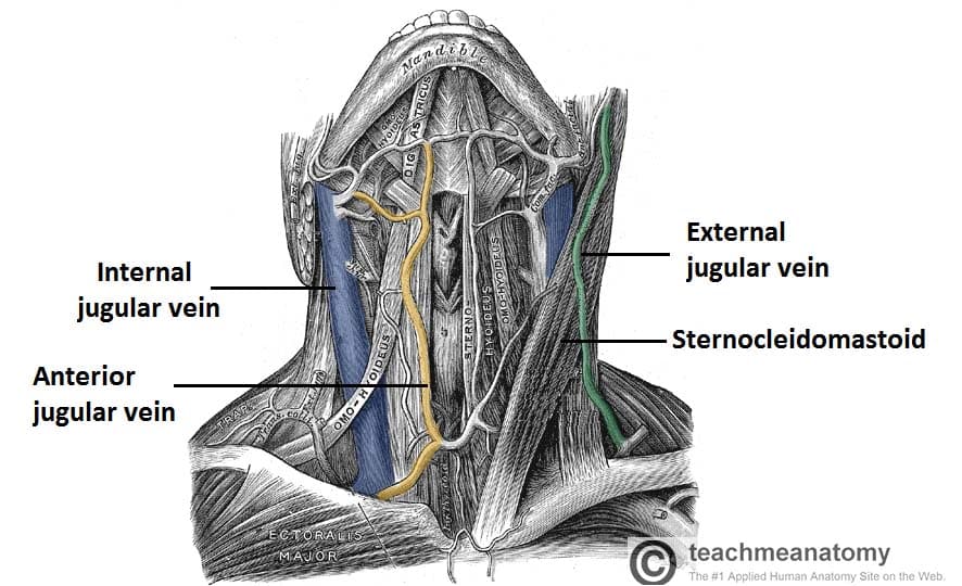
Structure and function. There are two sets of jugular veins: external and internal. The left and right external jugular veins drain into the subclavian veins.The internal jugular veins join with the subclavian veins more medially to form the brachiocephalic veins.Finally, the left and right brachiocephalic veins join to form the superior vena cava, which delivers deoxygenated blood to the.
Vena jugularis interna MedkoM
Percutaneous cannulation of the internal jugular vein uses anatomic landmarks to guide venipuncture and a Seldinger technique to thread a central venous catheter through the internal jugular vein and into the superior vena cava. Three approaches (central, anterior, and posterior) are used; the central approach is described here.
Anterior jugular vein Anatomy, tributaries, drainage Kenhub
Background. Internal jugular (IJ) vein thrombosis refers to an intraluminal thrombus occurring anywhere from the intracranial IJ vein to the junction of the IJ and the subclavian vein to form the brachiocephalic vein. It is an underdiagnosed condition that may occur as a complication of head and neck infections, surgery, central venous access.
Vena jugularis interna Anatomie und Klinik Kenhub
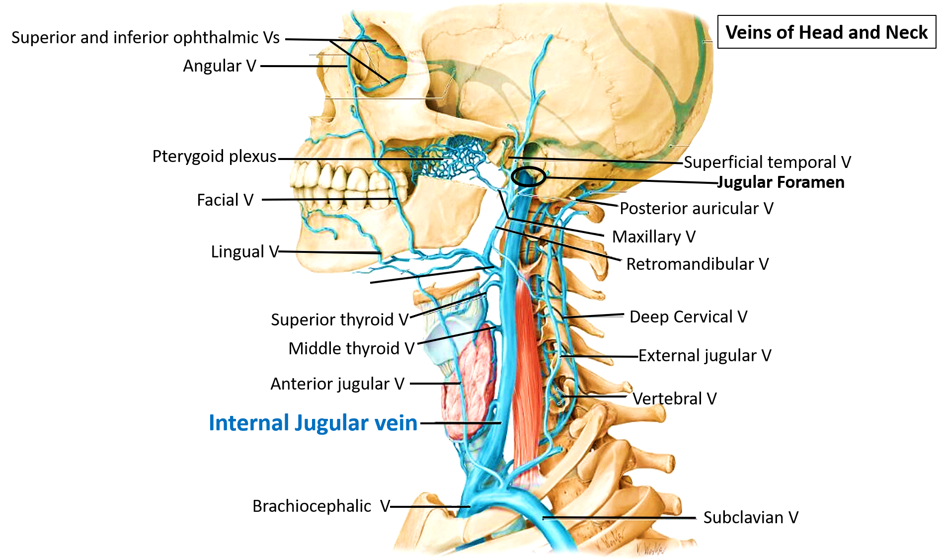
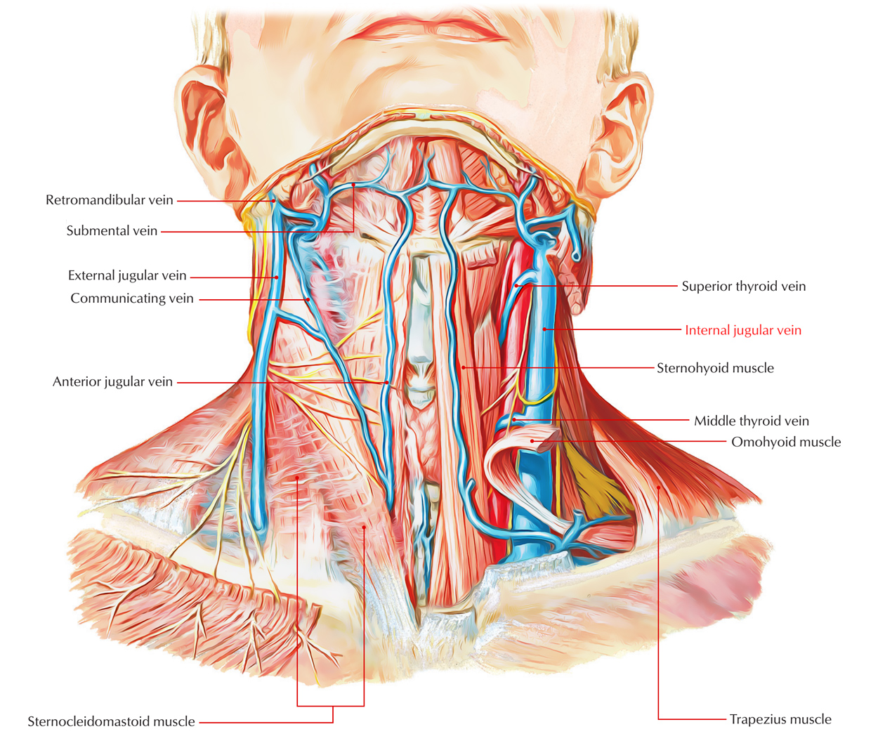
The internal jugular vein (IJV) is a paired vessel found within the carotid sheath on either side of the neck. It extends from the base of the skull to the sternal end of the clavicle. The internal jugular vein receives eight tributaries along its course.

Vena jugularis interna Wikipedia
The external jugular is a large vein used in prehospital medicine for venous access when the Paramedic is unable to find another peripheral vein [4] It is commonly used in cardiac arrest or other situations where the patient is unresponsive due to the pain associated with the procedure.

Internal Jugular Vein And Common Carotid Artery
Internal jugular (IJ) vein thrombosis refers to an intraluminal thrombus occurring anywhere from the intracranial IJ vein to the junction of the IJ and the subclavian vein to form the brachiocephalic vein. It is an underdiagnosed condition that may occur as a complication of head and neck infections, surgery, central venous access, local mali.
Internal Jugular Vein Commencement Termination Relations Tributaries Applied Anatomy
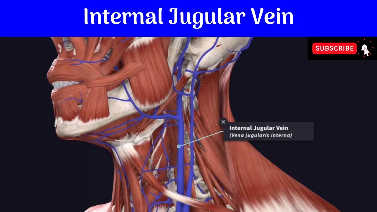
Function. Blood Flow. The internal jugular vein is the largest vein in the neck and is the main source of venous drainage, or blood flow, down from the brain, returning deoxygenated blood back from the head and neck to the heart, where it will be pumped to the lungs to become oxygenated again. The internal jugular vein also serves as the main.
Internal Jugular Vein Earth's Lab
The anterior jugular vein is a paired blood vessel that drains the anterior aspect of the neck. It emerges from the confluence of the superficial submandibular veins beneath the chin and drains into the external jugular vein. Less frequently, it may drain directly into the subclavian vein.

Anatomia do acesso venoso central
The internal jugular vein is a common route used by clinicians to access the central circulation for hemodynamical monitoring and stabilization due to its accessibility and anatomic location. Intravenous catheters cause injuries to the endothelium and vein wall inflammation.
Internal Jugular Vein Earth's Lab
The internal jugular vein is a paired jugular vein that collects blood from the brain and the superficial parts of the face and neck. This vein runs in the carotid sheath with the common carotid artery and vagus nerve. It begins in the posterior compartment of the jugular foramen, at the base of the skull.
Vena jugularis interna pacs
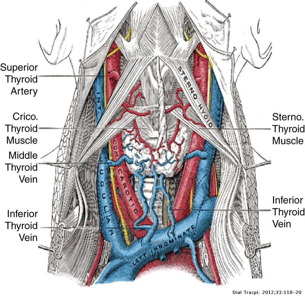
The internal jugular vein (IJV) originates at the jugular foramen, runs along the lateral neck, medially to the sternocleidomastoid muscle from the carotid triangle, and ends at the brachiocephalic vein. The IJV is one of the four components of the vascular sheath of the neck, together with the common and internal carotid arteries, the vagus.
Internal Jugular Vein—Central Venous Access Anesthesia Key
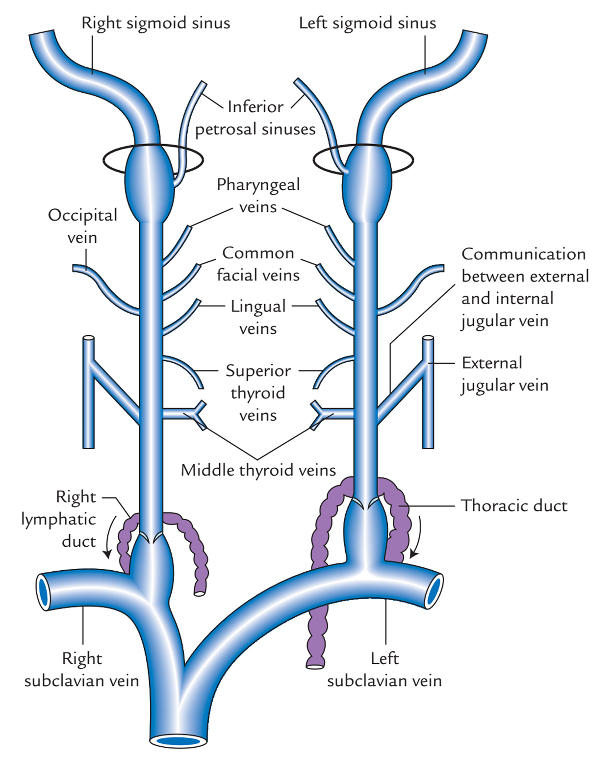
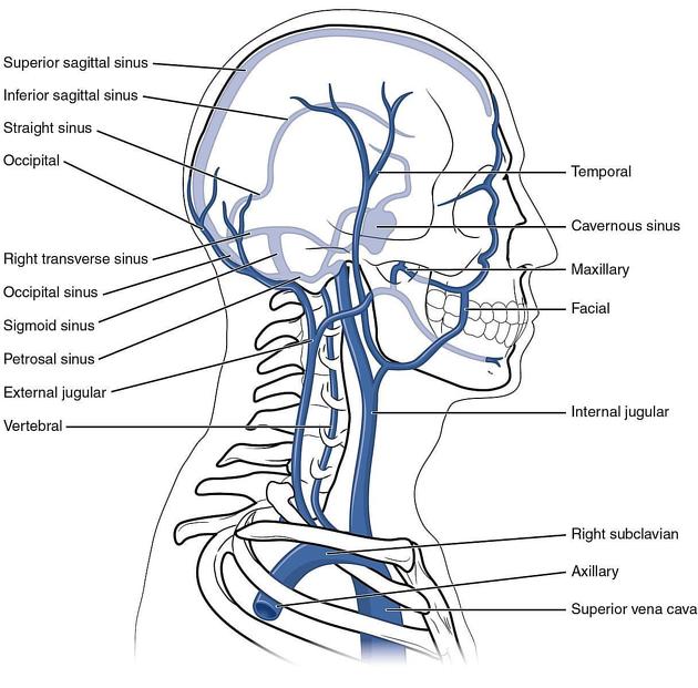
The internal jugular vein is formed by the union of the sigmoid and inferior petrosal venous sinuses. Here, the internal jugular vein is dilated as the superior bulb lying in the posterior part of the tympanic floor. Course
Internal Jugular Vein
The internal jugular vein (Latin: vena jugularis interna) is a blood vessel that arises from the junction of two intracranial venous sinuses - the inferior petrosal sinus and the sigmoid sinus.The internal jugular vein collects venous blood from the brain, skull, and superficial parts of the face and neck.. Internal jugular vein (lateral view) by Anatomy.app

Internal Jugular Vein Anatomy ANATOMY
Traditionally, when internal jugular vein cannulation has been performed, external anatomical landmarks and palpation have been used to guide insertion of the needle into the vessel. However,.
AAEM Resident and Student Association Anatomical Review of Jugular Central Line Placement
The internal jugular vein ( v. jugularis interna) collects the blood from the brain, from the superficial parts of the face, and from the neck. It is directly continuous with the transverse sinus, and begins in the posterior compartment of the jugular foramen, at the base of the skull.